
Imaging Facilities
Multispectral In Vivo Fluorescence Imaging
High-field (9.4 Tesla) Small Animal MRI (MicroMRI)
High-resolution small animal PET/CT (MicroPET/CT)
Dedicated Research Ultrasound Imaging (US)
High-frequency, High-resolution Ultrasound Imaging System
Multispectral In Vivo Fluorescence Imaging
A limiting factor for in vivo fluorescence imaging is the confounding
signal from the autofluorescence of the animal. Our multispectral imaging
system utilizes full multispectral imaging and spectral unmixing algorithms;
thus it is able to separate the tissue autofluorescence from the signal of interest,
thereby greatly increasing sensitivity and signal-to-noise. The multispectral in vivo
fluorescence imaging system could be used to guide the development and evaluation of new
drugs, nanoparticles, quantum dots, and imaging probes. Therefore, this device would provide
synergy among clinical research involving multiple emphasis areas: imaging, nanotechnology,
cancer, the drug discovery, inflammation/vaccines, and animal models of human disease.
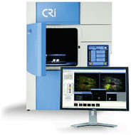
This system is located at the Quantitative BioImaging Laboratory (QBIL) in Wesley Woods Health Center, 2nd floor.
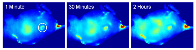
Fluorescence images of a tumor-bearing mouse after being injected with gold nanoparticle-conjugated photodynamic therapy drug at a) 1 min, b) 30 min, c) 120 min after intravenous tail injection. Unprecedented delivery efficiency and accumulation rate of the drug in the tumor is monitored via its fluorescence increase in the tumor area (white circle). Cheng Y, C Samia A, Meyers JD, Panagopoulos I, Fei B (co-corresponding author), Burda C. "Highly efficient drug delivery with gold nanoparticle vectors for in vivo photodynamic therapy of cancer". J Am Chem Soc. 2008 Aug 13;130(32):10643-7 Abstract|Full Text
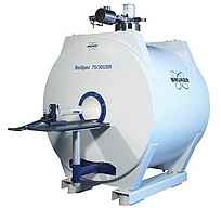
High-field (9.4 Tesla) Small Animal MRI (MicroMRI)
The multipurpose research system is a high-resolution magnetic resonance
spectroscopy and imaging scanner for 2- or 3-dimensional image reconstruction based on the principles
of nuclear magnetic resonance. The high-field system is equipped with multiple gradient sets specifically
for high-resolution in-vivo imaging applications (< 100um). The Biospec® system is also equipped with
broadband capabilities for multinuclear imaging and spectroscopy applications (ex. 1H, 31P, 15N, 13C, 19F).
The system is also capable of cardiac / respiratory gating / monitoring to limit the negative effects of
motion on image quality. This feature is critical for imaging animals with high respiration / heart rates.
This system is located at the Division of Animal Division (DAR) in the Whitehead Building at Emory University.
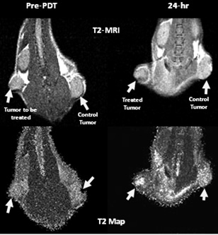
T2-weighted MR images of a tumor-bearing mouse before photodynamic therapy (PDT) and 24-hr after PDT (right). The signal intensity values changed 24-hr after the treatment. Compared to the T2 map pre-PDT (bottom), the T2 values increased 24-hr after the treatment, especially within the treated tumor (arrow). B Fei, H Wang, JD Meyers, D Feyes, NL Oleinick, JL Duerk, "High-Field Magnetic Resonance Imaging of the Response of Human Prostate Cancer to Pc 4-based Photodynamic Therapy in an Animal Model", Lasers in Surgery and Medicine, 39(9):723-30, 2007. Abstract|Full Text
High-resolution small animal PET/CT (MicroPET/CT)
Preclinical PET and CT imaging solutions provide insight into disease biology and evaluation of
novel therapeutic options in small animal models. Siemens Inveon is a revolutionary preclinical imaging platform,
providing integrated small animal PET and CT imaging and analysis. A brilliant combination of cutting-edge technologies
with multiple configuration options, Inveon is available as a fully combined system with single or multimodality configurations.
This system is located at the Center for Systems Imaging on the second floor of Wesley Woods Health Center Building.
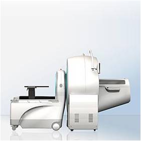
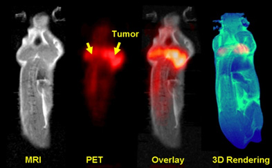
Figure 11. MicroPET and MR imaging for tumor response assessment in mice. High-resolution MRI and PET images are registered using our multimodality image registration software. After image fusion, the overlay image shows both MRI (gray) and microPET (red). The rendering image (blue from MRI and red from PET) shows both anatomical structure and metabolic information. B Fei, HS Wang, RF Muzic, CA Flask, DL Wilson, JL Duerk, DK Feyes, NL Oleinick, "Deformable and rigid registration of MRI and microPET images for photodynamic therapy of cancer in mice," Medical Physics, vol. 33, No. 3, pp. 753-760, 2006. Abstract|Full Text
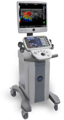
Dedicated Research Ultrasound Imaging (US)
The ultrasound imaging system (SonixTOUCH Research, Ultrasonix) is a full diagnostic ultrasound system with full research capabilities. This
means that the system can be used in a clinical environment to capture high quality diagnostic images, but also retrieve the raw data needed to
perform more in-depth analysis. Customized experiments can be performed by taking advantage of one of the many software toolkits that come
standard on the system, giving maximum flexibility to the user. Based on the OpenSONIX platform, the SonixTOUCH Research system provides an
open-architecture system that can be configured via software to operate in the various modes required for research. In addition, the research
interface not only allows you to capture data, but also to tweak up to 300 parameters that control how the ultrasound imaging and beamforming
is performed. The system is mobile and is used by the Quantitative BioImaging Laboratory in the Clinic B Building.
Data Acquisition
The SonixTOUCH Research system has the ability to acquire large cineloops of various types of data, ranging from raw RF signal in B-Mode,
Color, PW, 3D/4D modes or scan converted data, 8-bits or 16-bits envelope data. The system sequences can also be manually programmed and the
corresponding data acquired - for example to acquire full frames of pre-scan converted data in near-realtime.
OpenSONIX Platform
The SonixTOUCH Research system provides access (in investigational mode for use under IRB only) to the various ultrasound imaging parameters
to give the users the ability to alter sequences, processing and other features for their particular research use. Additional software plug-ins
can be developed in Matlab, Visual C++, communicate and synchronize with peripherals connected to the system.
Large Library of Tools
A dedicated Research user's group provides hundreds of posts with very helpful resources such as Matlab, Python, C Code to configure the system
and process the data.
High-frequency, High-resolution Ultrasound Imaging System
This VisualSonics Vevo® 2100 system with a new digital platform provides high-resolution (30 micron) ultrasound imaging for a wide range
of animal models and applications including cardiovascular, cancer and inflammation. The system expands the functionality, flexibility and
image quality of the Vevo 770 system, operating at frequencies with solid-state array transducers. The new MicroScan transducers provide
increased frame rates, superb contrast, unrivaled resolution and a wider field of view. The system is easy to use, non-invasive and fast,
providing extremely high throughput when needed. The frame rates in 2D are up to 740 fps (for a 4x4 mm field of view). It has color and Power
Doppler Modes for blood flow quantification & anatomical identification. The M-Mode single line acquisition allows high-temporal resolution
for LV functional analysis. It also includes the anatomical M-Mode for adjustable anatomical orientation in reconstructed M-Mode imaging,
3D-Mode Imaging & Volume Analysis, Nonlinear Contrast Imaging, and VevoStrain Analysis software for cardiac research.
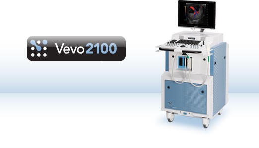
This system is located at the Emory Pediatric Research Center.