Label-free HSI for Surgical Margin Assessment
Quantification Tools for HSI During Surgical Guidance
Spectral-Spatial Classification Methods for Tumor Detection
Minimum Spanning Forest Based Method for HSI
Early detection of malignant lesions could improve both survival and quality of life of cancer patients. Hyperspectral imaging (HSI) has emerged as a powerful tool for noninvasive cancer detection and diagnosis, with the advantage of avoiding tissue biopsy and providing diagnostic signatures without the need of a contrast agent in real time. We developed a spectral-spatial classification method to distinguish cancer from normal tissue on hyperspectral images. We acquire hyperspectral reflectance images from 450 nm to 900 nm with a 2-nm increment from tumor-bearing mice. In our animal experiments, the hyperspectral imaging and classification method achieved a sensitivity of 93.7% and a specificity of 91.3%. The preliminary study demonstrated that HSI has the potential to be applied in vivo for noninvasive detection of tumors.
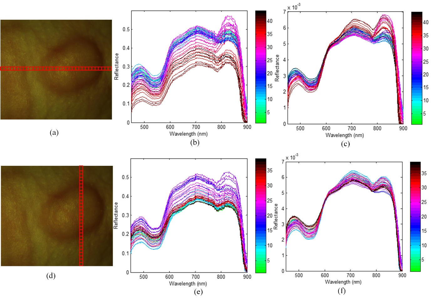
Effects of Pre-processing on Spectra

Effects of the pre-processing on spectra as selected from different regions of a mouse image. (a) and (d) are the same ROI covering the tumor area; the horizontal and vertical locations are composed of square areas of "10×10" pixels. (b) and (c) shows the average spectra of each square from left to right before and after pre-processing. (e) and (f) shows the average spectra of each square from top to bottom before and after pre-processing.
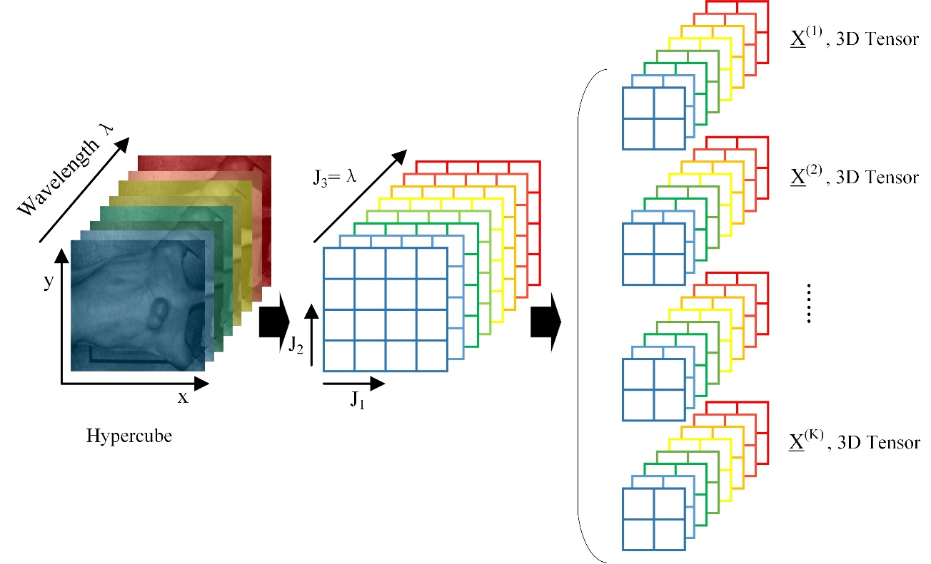
Spectral-Spatial Tensor

Spectral-spatial tensor representation of hypercube. Image stacks on the left is the hypercube of a tumor-bearing mouse. Images stacks in the middle shows that a hypercube (J_1×J_2×J_3) can be divided into small patches. Image stacks on the right shows that each pixel inside a hypercube can be represented by a small patch centered at that pixel. This patch containing information from both the pixel and its neighborhood can be represented in a mathematical form of 3D tensor.
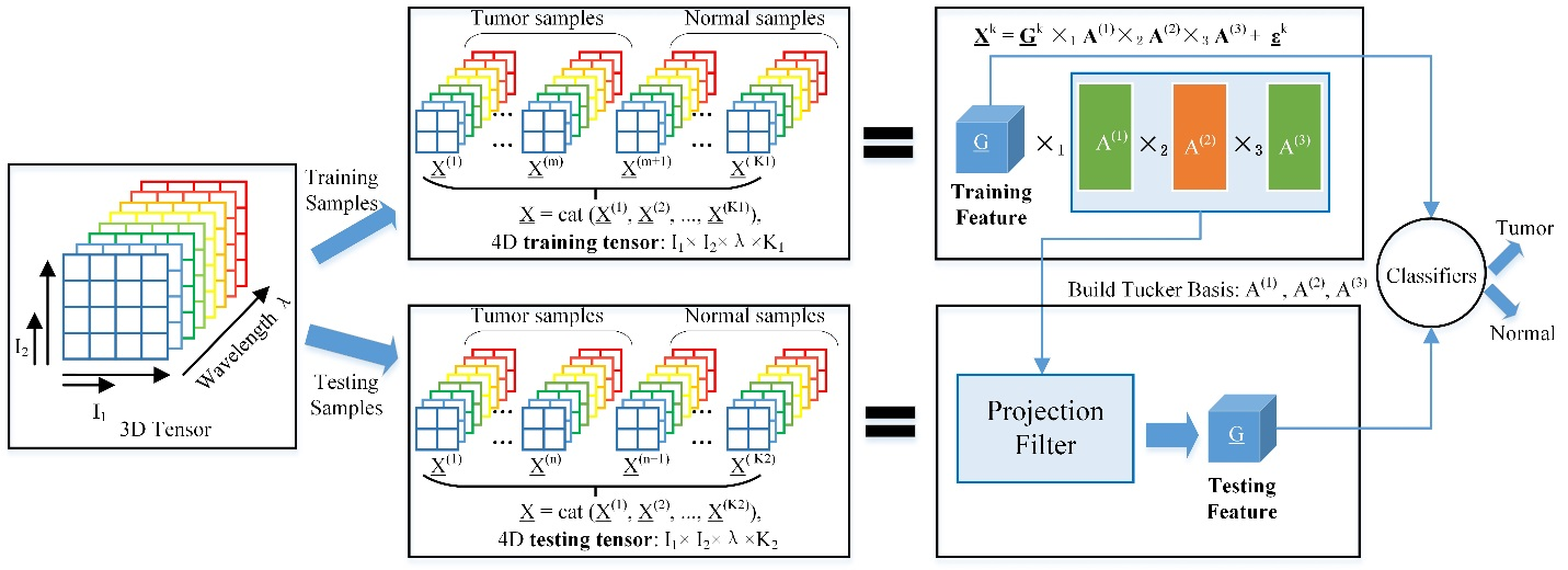
Tensor Decomposition

Tensor decomposition and feature extraction for the classification of tumor and normal tissue.
