Multiparametric MRI for the Prostate
PET/CT Imaging of Prostate Cancer
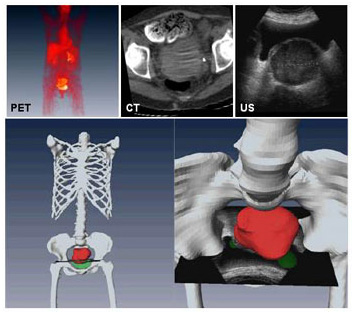
PET/CT/Ultrasound Fusion of Gynecological Cancer
Combined CT, PET and ultrasound images could help diagnose gynecological cancers
A method of automatically combining CT, PET, and ultrasound scans into one image may help clinicians diagnose gynecological cancers. Together, the three modalities provide a clearer picture of indeterminate solid masses in the pelvic area.
Ultrasound is the gold standard for examining abnormal pelvic masses to differentiate between a cyst and a solid mass. It cannot differentiate, however, between a benign solid mass and a malignant one. PET scans showing metabolism and blood flow within an area can provide more information about malignancy, but localizing the pathology can be a problem without CT. Putting PET/CT and ultrasound scans together yields an image with the benefits of both.
Dr. Baowei Fei and colleagues described their method of adding ultrasound to an already-fused PET/CT image in a paper presented at the 2007 American Institute of Ultrasound in Medicine meeting last month in New York City.
By registering ultrasound with CT, the researchers automatically registered the ultrasound with the preregistered PET scan as well, providing a level of functional imaging over the two sources of anatomic imaging. The researchers used a slice-to-volume registration method previously applied to MR images.
"As each image may have thousands or millions of pixels, the intensity values of the pixels represent the information of the image. We use the rich information from the intensity values to compute the mutual information. If two images are registered, their mutual information value is maximized. ... This method is fully automatic and does not need landmarks for the registration," Fei said.
The computations take only seconds on a desktop computer, and Fei thinks real-time registration could be done with powerful enough hardware.
The researchers tested their approach in 100 simulated trials, using real clinical data with variations in noise levels and image contrast. They had a success rate of 98% with a mean error of less than 0.1 mm for translation and 0.1° rotation. They also tested the technique on a cervical cancer patient, and visual inspection indicated excellent registration. Since publishing the paper, the researchers have tried their procedure on additional patient data and anatomic targets such as the prostate.
"The method will provide a powerful tool for clinical applications," Fei said. "Combining ultrasound and PET/CT images has great potential to detect cancer at an early stage, improve the sensitivity and specificity, and better diagnose normal and malignant tissues."
He also looked forward to its use with image-guided radiation therapy and image-guided radiofrequency ablation.
Fei's colleagues contributing to the study included Drs. Nami Azar, Peter Faulhaber, Paul Rochon and Dean Nakamoto.
Fusion of PET, CT and Ultrasound

Fusion of PET, CT, and ultrasound images for improved diagnosis of gynecological cancer. Top: PET, CT, and ultrasound images of same patient. Bottom: Three-D fusion of three imaging modalities. Combined PET/CT provides both anatomic (bone in white) and pathologic (tumor in red) information. At right, ultrasound image registered with PET/CT demonstrates malignancy of mass in pelvic area.
