PET/Ultrasound Fusion Targeted Biopsy of Prostate Cancer
Image-guided Focal Therapy of Prostate Cancer
Label-free HSI for Surgical Margin Assessment
Photodynamic Therapy of Head and Neck Cancer
One in six men will be diagnosed with cancer of the prostate during their lifetime. Systematic transrectal ultrasound (TRUS)-guided prostate biopsy is considered the standard method for prostate cancer detection. Current biopsy techniques have a significant sampling error and a low sensitivity (27%-54%) and can miss up to 30% of cancers. As a result, a patient may have a “negative” biopsy but may, in fact, be harboring an occult cancer. Alternatively, a diagnosis of cancer may have been made, but the patient is under-staged because the most aggressive histologic region of the tumor has not been sampled. Because of these limitations, patient management may not be optimized. In many men, current diagnostics are insufficient to distinguish aggressive prostate cancer, which requires treatment, from the non-aggressive disease, which may be well-managed with active surveillance. In an era of focal therapy/prostate sparing treatments for small volume prostate cancer, it is imperative to be able to precisely sample the treated area to detect whether the treatment was effective.
To improve the cancer detection rate from the current standard of care practice with an initial prostate biopsy, the development of new targeted biopsy technology is an active research area. MR/ultrasound fusion guided biopsy represents one approach. It has been reported that MR/TRUS fusion biopsy detects more cancer per core than standard 12-core TRUS biopsy. The use of three-dimensional (3D) tracking and image fusion has the potential to improve current methods for diagnosis and follow-up of prostate cancer.
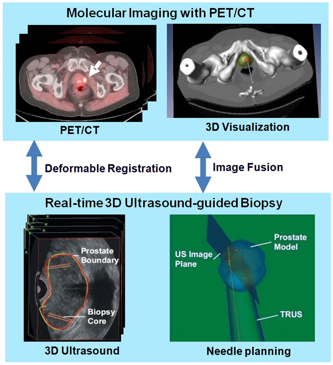
Positron emission tomography (PET) can detect metabolic and functional information of cancer. PET imaging agents have been developed for prostate cancer detection and staging. PET imaging with new molecular imaging tracers has shown promising results for detecting and localizing prostate cancer in humans. PET/CT may show focal uptake at the tumor and thus can provide location information to direct targeted biopsy of the prostate. By combining PET/CT with 3D ultrasound images, multimodality image-guided targeted biopsy has become a promising technology for improved detection and diagnosis of prostate cancer.

Molecular image-directed, 3D ultrasound-guided biopsy. Top: The PET/CT images with 18F-FACBC were acquired from a prostate patient. PET/CT images show a focal lesion within the prostate (white arrow). The 3D visualization of the pelvis and the prostate can be used to aid the insertion of the biopsy needle into a suspicious tumor target. Bottom: During biopsy, a mechanically assisted navigation device was used to acquire 3D TRUS images from patients. The prostate boundaries on TRUS images were segmented and were used to generate a 3D model of the prostate. The 3D prostate model and real-time TRUS images are used to guide the biopsy in patients.

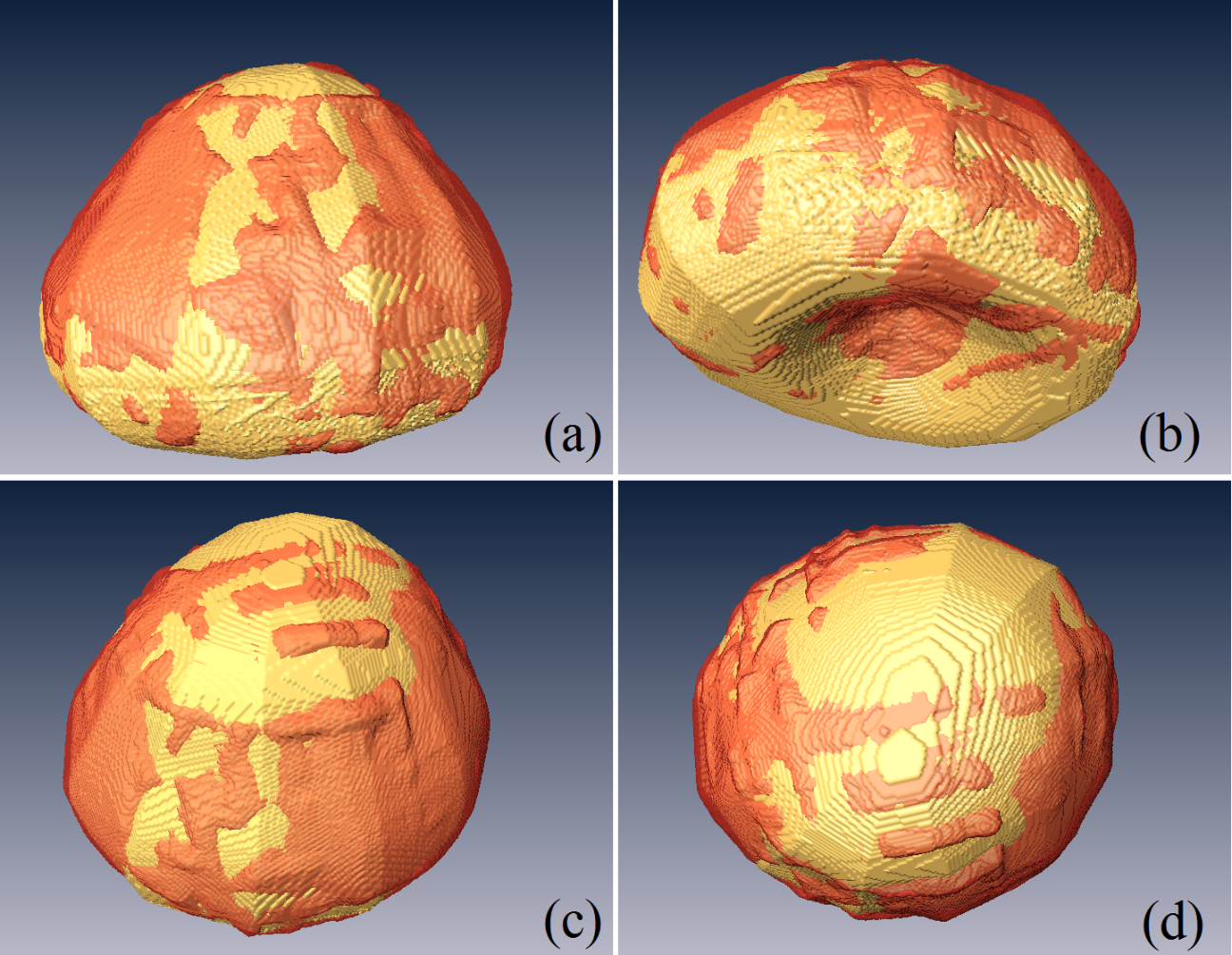
Three-dimensional visualization of the segmented prostate (red) as compared to the manual gold standard (gold color) in four different views (a-d) of the same human prostate.
For more information about the targeted biopsy study, please visit: https://www.clinicaltrials.gov/ct2/show/NCT02744534/
