
Image Registration
• Thin-plate spline (TPS) deformable image registration
• B-spline deformable image registration
• Finite element model-based deformable registration
For soft tissue, e.g. tumor, registration, we developed a finite element model (FEM)-based deformable registration method. We have applied this FEM registration method to tumor MRI and PET images. In the first step, we applied a rigid registration algorithm to align the cropped MRI and PET images using three translations and three rotations. After registration, we manually segmented the tumor slice-by-slice on both high-resolution MRI and PET image volumes. We then applied the deformable registration algorithm. The registration approach deforms the tumor surface from the MRI volume toward that from the PET image. The displacements at the surface vertices are the force that drives the elastic surface from MRI toward that from the PET image. The tumor was modeled as a linear isotopic elastic material. The FEM model was used to infer volumetric deformation of the tumor from the surface. The force is then integrated over each element and is distributed over the nodes belonging to the element using its shape functions. After obtaining the displacement field for all vertices, we used a linear interpolation to obtain the deformed image volume of the tumor.
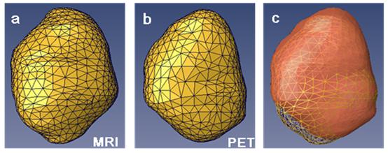
Figure 3. Three-dimensional meshes of a tumor. (a) Tumor segmented from a high-resolution MR volume. (b) Same tumor from the corresponding microPET emission images. (c) Color overly of the tumor from MRI (yellow) and PET (red). The tumor deformed during the two imaging sessions.
• Two-dimensional (2D) image registration software• Three-dimensinal (3D) image registration software
• 3D to 2D Registration
• Slice to Volume Registration
Image Classification
Image Segmentation
Attenuation Correction
Partial Volume Correction
Motion Correction