
Image Registration
• Thin-plate spline (TPS) deformable image registration
We created and evaluated an almost fully automated, 3D non-rigid registration algorithm using mutual information and a thin-plate spline (TPS) transformation for MR images of the prostate and pelvis. In the first step, an automatic rigid-body registration with special features was used to capture the global transformation. In the second step, local feature points were registered. An operator entered only five feature points (FPs) located at the prostate center, in the left and right hip joints, and in the left and right distal femurs. The program automatically determined and optimized other FPs on the external pelvic skin surface and along the femurs. More than 600 control points were used to establish a TPS transformation for deformation of the pelvic region and the prostate. Ten volume pairs were acquired from three volunteers in the diagnostic (supine) and treatment positions (supine with legs raised). Various visualization techniques showed that warping rectified the significant pelvic misalignment caused by the rigid body method. Gray-value measures of registration quality, including mutual information, correlation coefficient, and intensity difference, all improved with warping. The distance between prostate 3D centroids was 0.7 ± 0.2 mm following warping compared to 4.9 ± 3.4 mm with rigid-body registration. The semiautomatic non-rigid registration works better than rigid body registration when the patient position is changes significantly between acquisitions; it could be a useful tool for many applications of prostate diagnosis and therapy.
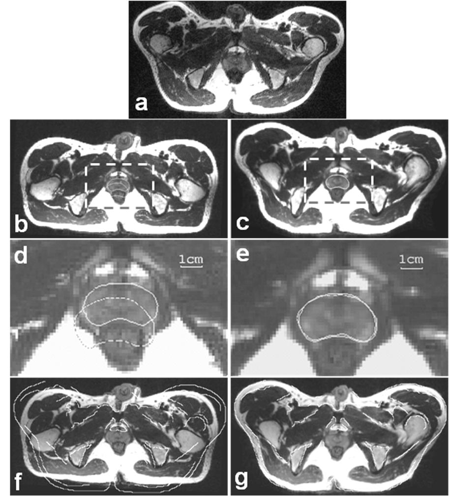
Figure 1. Comparison of non-rigid and rigid-body registration for volumes acquired in the treatment and diagnostic positions. Image (a) is from the reference volume acquired in the treatment position and where the prostate is manually segmented. Images in the left and right columns are from the floating volume acquired in the diagnostic position following rigid-body and non-rigid registration, respectively. To show potential mismatch, the pros tate contour from the reference in (a) is copied to both (b) and (c) and is magnified as the dashed contours in (d) and (e). The movement of the prostate to the posterior is corrected with warping (e) but not with rigid-body registration (d). Pelvic boundaries manually segmented from the reference show significant misalignment with the rigid body (f) that is greatly improved with warping (g). All registration experiments are performed in three-dimensions (3D). Transverse slices covering the prostate are selected from the 3D image volumes.
• B-spline deformable image registration• Finite element model-based deformable registration
• Two-dimensional (2D) image registration software
• Three-dimensinal (3D) image registration software
• 3D to 2D Registration
• Slice to Volume Registration
Image Classification
Image Segmentation
Attenuation Correction
Partial Volume Correction
Motion Correction