Multiparametric MRI for the Prostate
PET/CT Imaging of Prostate Cancer
PET/CT/Ultrasound Fusion of Gynecological Cancer
PET imaging plays a central role in the detection of recurrent prostate carcinoma in the prostate bed and in the differentiation of prostatic from extraprostatic recurrence. Conventional methodology including computed tomography (CT), magnetic resonance imaging (MR), transrectal ultrasound, bone scan and ProstaScint SPECT. Various PET imaging agents have been developed for prostate cancer detection and staging. These include radiolabeled choline, fluorocholine, acetate, minibody, FPA, FDHT, J591, methionine, DCFBC, and etc. FACBC (Axumin, Blue Earth Diagnostics) is indicated for diagnosis of recurrent prostate cancer in men who have elevated blood levels of prostate specific antigen (PSA) after previous treatment, which was originally developed at Emory University and was recently approved by the by the U.S. Food and Drug Administration.
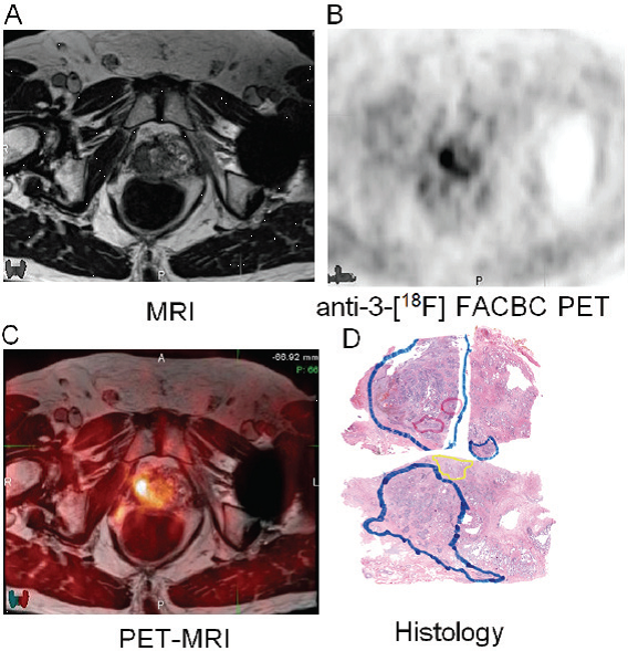
MRI (A), FACBC PET acquired at 28 min post radiotracer administration (B), co-registered PET-MRI (C) and histology (D). In this patient uptake in the right anterior and posterior mid sextants correlate with presence of tumor while a small tumor focus in the left anterior sextant is not visualized. On the histology, solid blue represent Gleason 4, solid red represents Gleason 5, and yellow represents BPH. (Schuster DM, Taleghani PA, Nieh PT, Master VA, Amzat R, Savir-Baruch B, Halkar RK, Fox T, Osunkoya AO, Moreno CS, Nye JA, Yu W, Fei B, Wang Z, Chen Z, Goodman MM. Am J Nucl Med Mol Imaging. 2013;3(1):85-96)