Multiparametric MRI for the Prostate
PET/CT Imaging of Prostate Cancer
PET/CT/Ultrasound Fusion of Gynecological Cancer
Ultrasound is the gold standard for examining abnormal pelvic masses to differentiate between a cyst and a solid mass. It cannot differentiate, however, between a benign solid mass and a malignant one. PET scans showing metabolism and blood flow within an area can provide more information about malignancy, but localizing the pathology can be a problem without CT. Putting PET/CT and ultrasound scans together yields an image with the benefits of both.
A method of automatically combining CT, PET, and ultrasound scans into one image may help clinicians diagnose gynecological cancers. Together, the three modalities provide a clearer picture of indeterminate solid masses in the pelvic area. By registering ultrasound with CT, we automatically registered the ultrasound with the preregistered PET scan as well, providing a level of functional imaging over the two sources of anatomic imaging. We used a slice-to-volume registration method previously applied to MR images for this application.
Fusion of PET, CT and Ultrasound
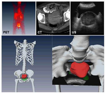
Fusion of PET, CT, and ultrasound images for improved diagnosis of gynecological cancer. Top: PET, CT, and ultrasound images of same patient. Bottom: Three-D fusion of three imaging modalities. Combined PET/CT provides both anatomic (bone in white) and pathologic (tumor in red) information. At right, ultrasound image registered with PET/CT demonstrates malignancy of mass in pelvic area.