High-frequency Ultrasound Imaging for Cardiac Fibers
3D Ultrasound and MR Diffusion Tensor Imaging
Simulated Ultrasound Image from MR Diffusion Tensor Image
Cardiac Fibers in Pig Hearts
Cardiac ultrasound simulation can have important applications in the design of ultrasound systems, understanding the interaction effect between ultrasound and tissue, and setting the ground truth for validating quantification methods. Current ultrasound simulation methods fail to simulate the myocardial intensity anisotropies. The proposed cardiac ultrasound image simulation method is based on diffusion tensor imaging (DTI) data of the heart. The method utilizes both the cardiac geometry and the fiber orientation information to simulate the anisotropic intensities in B-mode ultrasound images. Before the simulation procedure, the geometry and fiber orientations of the heart are obtained from high-resolution structural MRI and DTI data, respectively. The simulation includes two important steps. First, the backscatter coefficients of the point scatterers inside the myocardium are processed according to the fiber orientations using an anisotropic model. Second, the cardiac ultrasound images are simulated with anisotropic myocardial intensities. The proposed method was also compared with two other non-anisotropic intensity methods using 50 B-mode ultrasound image volumes of five different rat hearts. The simulated images were also compared with the ultrasound images of a diseased rat heart in vivo. A new segmental evaluation method is proposed to validate the simulation results. The average relative errors (AREs) of five parameters, i.e. mean intensity, Rayleigh distribution parameter σ, and first, second and third quartiles, were utilized as the evaluation metrics. The simulated images were quantitatively compared with real ultrasound images in both ex vivo and in vivo experiments. Our proposed ultrasound image simulation method can realistically simulate cardiac ultrasound images of the heart using high-resolution MR-DTI data. The proposed simulation method uses MR-DTI data and realistically generates ardiac ultrasound images with anisotropic intensities inside the myocardium. The ultrasound simulation method could provide a tool for many potential research and clinical applications in cardiac ultrasound imaging.
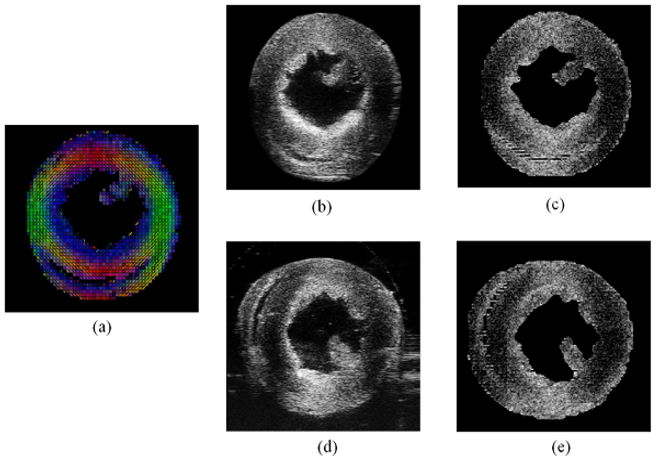
Simulated ultrasound images from both orthogonal imaging directions of the same rat heart. (a) Cardiac fiber orientations from DTI. (b) Real ultrasound image with vertical beam direction. (c) Simulated result of (b). (d) Real ultrasound image with horizontal beam direction. (e) Simulated result of (d).
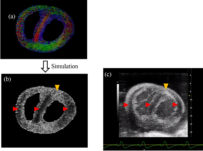
Comparison between the simulated myocardial ultrasound image and the in vivo acquired one of a diseased rat heart with pulmonary artery hypertension. (a) Fiber orientations of the heart from DTI ex vivo. (b) Simulated ultrasound image based on DTI data. (c) Acquired ultrasound image in vivo. The red arrows indicate the lower intensity regions of myocardium and the yellow ones indicate the higher intensity regions, which are affected by different fiber orientations.
Fiber Modeling and Ultrasound
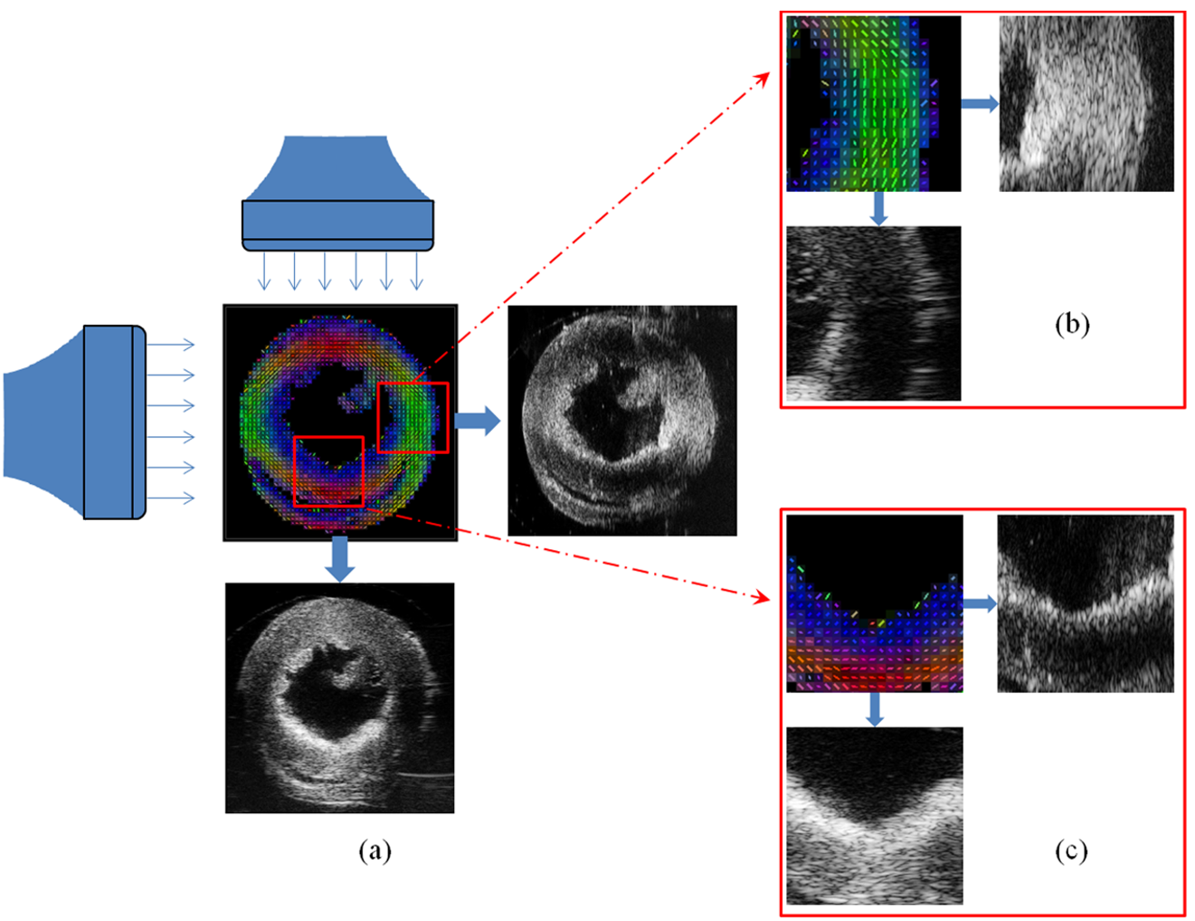
Relationship between ultrasound intensity and cardiac fiber orientations. (a) Two ultrasound images acquired from both orthogonal directions to image one heart, where middle is the distribution of its fiber orientations. (b) Magnified heart region with main vertical fiber orientations and both corresponding ultrasound images from orthogonal imaging directions. (c) Magnified heart region with main horizontal fiber orientations and both corresponding ultrasound images from orthogonal imaging directions. The enlarged regions in both (b) and (c) demonstrate how different angles between cardiac fiber orientations and ultrasound beam directions affect the ultrasound intensities.
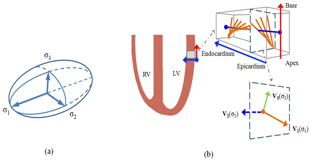
Relationships among ellipsoid model, DTI eigenvectors, and cardiac fiber orientations. (a) Ellipsoid model, where σ1 > σ2 > σ3. (b) Illustrated relationship between three DTI eigenvectors and fiber microstructures: primary eigenvector corresponding to σ1 indicates the fiber orientation, secondary eigenvector corresponding to σ2 indicates the sheet direction, and tertiary eigenvector corresponding to σ3 indicates the sheet norm direction.
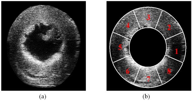
Illustration of the selected eight segments for the evaluation of ultrasound simulations. (a) Ultrasound image of a rat heart. (b) Divided eight segments of (a), where the white lines indicate the boundaries of the segments.
Virtual Phantoms
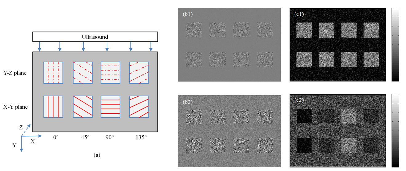
Ultrasound simulations of a virtual fiber phantom with different fiber orientations. (a) Illustration of the fiber phantom, where the top row indicates the various fiber orientations in the Y-Z plane and the bottom row indicates the ones in the X-Y plane. (b1) Point scatterers randomly distributed. (b2) Point scatterers filtered based on the fiber orientations. (c1) Ultrasound simulation of (b1), showing similar intensities for the eight objects. (c2) Ultrasound simulation of (b2), showing different intensities for the objects with different fiber orientations.
Simulation Results
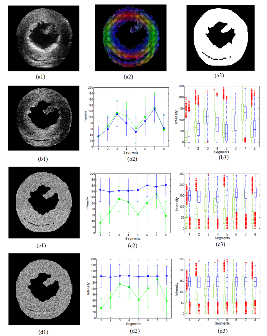
Comparisons between real cardiac ultrasound images and the simulation results of three different methods. (a1) Real ultrasound image. (a2) Corresponding cardiac fiber orientations from DTI. (a3) Corresponding myocardial geometry from the structure MR. (b1) Simulated ultrasound image with the scatterers filtered by the DTI-derived orientations (M1). (b2-b3) Mean, median and quartile intensity comparisons of the eight segments between (a1) and (b1), respectively. (c1) Simulated ultrasound image with the scatterers randomly distributed (M2). (c2-c3) Mean, median and quartile intensity comparisons of the eight segments between (a1) and (c1), respectively. (d1) Simulated ultrasound image with the scatterers filtered by the random orientations (M3). (d2-d3) Mean, median and quartile intensity comparisons of the eight segments between (a1) and (d1), respectively. Green lines indicate the results of the real image and blue lines indicate the results of the simulated images.
Human Heart Simulation
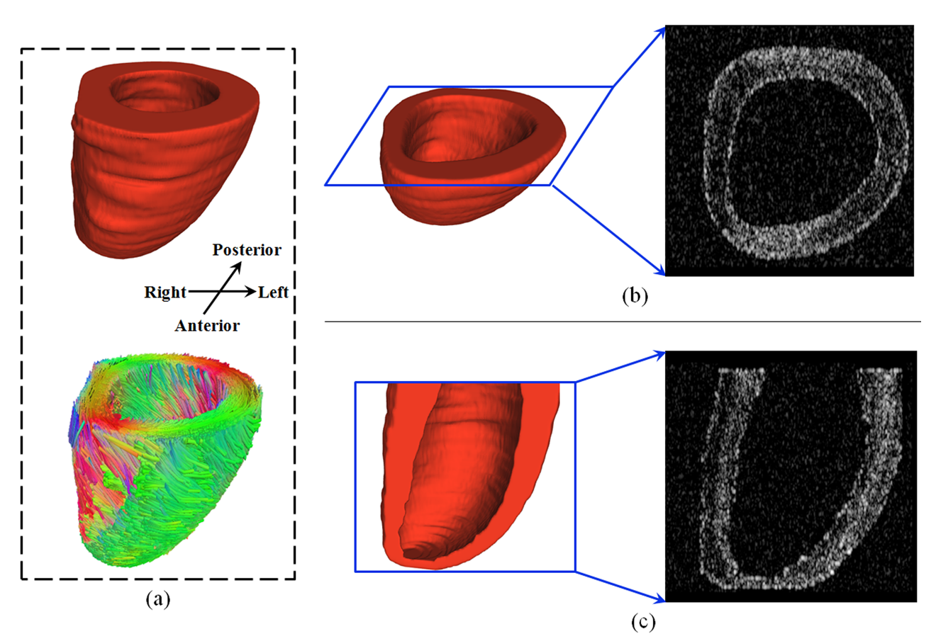
Simulated myocardial ultrasound images of the left ventricle of a human heart. (a) Architecture of the human heart: The upper row is the geometry from structure MR and the lower row is the fiber orientations from DTI. (b) Simulated ultrasound image from the short axis view. (c) Simulated ultrasound image from the long axis view.
